Monday, November 23, 2020

Other Common Musculoskeletal Conditions

 Tag: Muscuoskeletal Conditions

Other Common Musculoskeletal Conditions Treated In Delhi


Tennis/Golfer’s Elbow (Lateral/Medial Epicondylitis)

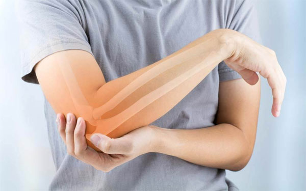
These are common problems involving staining/overuse of forearm tendons which attach to the bony prominences on the upper arm bone close to the elbow joint. Tennis elbow involves the tendons attaching to the outer side of elbow and repetitive activities involving gripping or twisting of forearm makes this pain worse. It may affect one or more tendons and it’s not uncommon for the pain to radiate down further into the forearm. Golfers elbow involves the tendons on the inner side of elbow.

Treatment involves activity modification, physiotherapy and painkillers. In clinic diagnostic ultrasound scan followed by PRP or steroid injection and physiotherapy can help effectively deal with the condition.

Achilles Tendinopathy

Achilles tendon attaches the calf muscles to the heel bone and is the biggest tendon in the body. It is located behind and above the heel. Overuse or underuse of the tendon can predispose to tendinopathy hence it may be seen in very active people or in those with a sedentary life style. Other predisposing factors include incorrect footwear, poor flexibility, being overweight and sudden increase in physical activities.

Most common symptom is pain behind or just above the ankle with associated stiffness and swelling. Diagnosis can be made with the help of an ultrasound scan. Conservative management involves rest, activity and footwear modification, weight management, medications and physical therapy. Tendon sheath PRP or steroids injections under ultrasound guidance, extracorporeal shockwave therapy are reserved for those not responding to conservative management.

Plantar Fasciitis

Plantar Fascia is a tissue band in the sole of foot which stretches from the front part of heel to the toes. It works like a shock absorber playing an important role in pushing the foot off from the ground during walking. Repeated stress/ injury to the fascia can lead to a condition called plantar fasciitis, which present as pain on the underside of heel worse on taking first few steps after a period of rest. Risk factors include using footwear with poor cushioning, being overweight, sudden change in activity levels, and calf muscle tightness.

The diagnosis is made by history, clinical examination and is aided by an ultrasound scan. Ultrasound can help in identifying any partial tears, fascia rupture and any foci of calcification or calcaneal spurs. An x-ray may be requested to rule out any bony spurs.

Conservative management involves

  • Using correct footwear with shock absorbing sole and arch support
  • Rest/ activity modification
  • Physiotherapy involving stretching of plantar fascia and achilles tendon
  • Night splints
  • Painkillers
  • Weight management and
  • Ultrasound guided injections - Platelet Rich Plasma (PRP) or steroid are reserved for patients with a poor response to conservative management.

De Quervain’s tenosynovitis

This condition results from irritation of two tendons as they travel in close proximity to each other in their course from the wrist towards the thumb. Irritation results in inflammation, swelling and thickening of the tendons or their covering sheath impacting their ability to glide freely during wrist and thumb movements. It is seen more commonly in women and presents as pain, swelling at the base of thumb, wrist.

Treatment involves rest, splints, medications, physiotherapy and if not responding to conservative measures then steroid injection. Ultrasound scans can help in verifying the diagnosis and precise injection of steroid to reduce the pain.

Injections for proximal hamstring tendinopathy & ischial bursitis

Hamstrings are a group of muscles present at the back of thigh. They extend from the pelvis (ischial tuberosity) to the knee and play an important role is everyday activities such as bending or running. Muscles attach to the bones with the help of a special type of tissue called tendons. With overuse, misuse or injury, these tendons can get inflamed, torn leading to development of a condition called tendinopathy. Involvement of proximal part of hamstrings presents as buttock pain radiating down the back of knee. Pain may be worse on sitting on a firm seat. Sciatic nerve is present close by and its irritation can cause pain to radiate further down the leg.

Condition such as unequal leg length, core and pelvic muscle weakness, being overweight and repeated overloading with insufficient warm up predispose to development of hamstring tendinopathy. Higher incidence is seen in runners, football players, dancers and older adults who do a lot of walking.

Treatment options include rest, activity modification, physical therapy and medications. If these fail to produce desired results then injections with Platelet Rich Plasma (PRP), autologous blood (ABI) or steroids are considered. Percutaneous tenotomy is another option. Injections are performed under ultrasound guidance in the peritendinous region. Direct injection into the tendons is avoided. These are performed under local anaesthesia as an outpatient procedure or a day case.

Platelet Rich Plasma (PRP)

Platelets are one of the blood components. They help in clotting and contain growth factors which promote the healing process. PRP is a blood plasma with concentrated platelets. PRP therapy is an attempt to utilise body’s natural ability to heal itself. It is utilised for tendon or ligament injuries which have not responded to conservative measures such as tennis elbow, golfer’s elbow, plantar fasciitis, achilles tendinosis.

The procedure involves collecting a blood sample from the patient which is then placed in a spinning machine to separate different blood components. The component containing high platelets is separated and then injected at the intended site under ultrasound guidance. Most people require 1-3 injections at 4 weekly intervals depending on the response. Growth factors are released from the platelets influence the process and accelerate the repair of tendon or ligaments.

Trigger Point Injections

Muscles ability to contract and relax plays an important role in body functioning. When muscles fail to relax, they form knots or tight bands known as trigger points. In simple words trigger points are irritable areas/ bands of tightness in a muscle. Pressure over a trigger point produces local soreness and may refer pain to other body parts. Common causes include inflammation, injury of the muscle or the neighbouring structures. Poor posture and repetitive strain are other predisposing factors. Trigger points can limit the range of movement; affect posture predisposing other areas to unaccustomed strain. They are more commonly observed in head, neck, and shoulder muscles.

Trigger point injections are performed in an outpatient/ day-care setting and the procedure involves injecting local anaesthetic with or without a small dose of steroid into the painful muscle. The local anaesthetic blocks the pain sensations and the steroids help in reducing the inflammation, swelling. I prefer to perform these injections under ultrasound guidance as it improves the accuracy and reduces the chances of complications. Post injection physiotherapy is essential to prevent recurrence and maximise the benefits.

Read More Blog:

Saturday, November 7, 2020

Complex Regional Pain Syndrome (CRPS)


Complex Regional Pain Syndrome (CRPS)


 Complex Regional Pain Syndrome (CRPS) is one of the causes of persisting limb pain. It can occur after surgery or injury and sometimes the injury is so trivial that you may not even remember it. In other cases the injury may be more severe with or without nerve damage. The pain however lasts much longer and is more severe than expected. It can range from mild self-limiting to chronic debilitating condition affecting activities of daily living and quality of life. The reason why CRPS develops is unclear and multiple mechanisms are thought to be involved. A combination of signs and symptoms is used to make the diagnosis as there is no specific diagnostic test. Investigations are helpful to exclude other conditions such as infection and rheumatologic conditions which may have similar presentation.


Pain in CRPS is accompanied by other signs and symptoms such as

  • Hypersensitivity of the affected limb
  • Swelling, abnormal sweating
  • Involved limb may feel unusually cold or warm with or without colour changes
  • Nail, skin and hair changes
  • Stiffness, weakness, abnormal tone of muscles and jerky movements

Management

The aim in CRPS management is pain control and functional recovery. Early diagnosis and treatment using multi- disciplinary approach is preferred. Your pain management specialist may recommend interventions such as intravenous drug infusions, sympathetic blocks such as stellate ganglion block or lumbar sympathetic blocks and neuromodulation. These are used in combination with medications, physiotherapy and psychology input.  

Medications prescribed depend on the phase of disease and the predominant symptoms. A combination of anti inflammatory drugs, neuropathic medications and opioids is commonly utilised. Some other medications including bisphosphonates, free radical scavengers (topical 50% dimethylsulfoxide- DMSO), oral steroids etc. may also be used.

Your physiotherapist will teach you desensitisation techniques if the limb sensitivity is increased. It helps in preventing problems due to weakening and reduced usage of the affected limb. Strengthening exercises are incorporated as the pain is adequately controlled. He may consider specialist interventions such as mirror therapy or graded motor imagery if indicated.

Read Another Blog

Shoulder Pain Treament In Gurgaon

Cancer Pain Treament In Gurgaon

Monday, November 2, 2020

Elbowing the tennis elbow pain out of your life – Information Leaflet on Tennis Elbow

 Tags >> causes the tennis elbowsymptoms of tennis elbow,  Tennis Elbow Treatment in DelhiPain Management Centre in Delhipain specialist doctor in Delhi

Elbowing the tennis elbow pain out of your life – Information Leaflet on Tennis Elbow

This leaflet has been written to help you understand more about your elbow problem. It is not intended to be a substitute for professional medical advice and should be used in conjunction with the information provided by your treating doctor.

What is tennis elbow?

Tennis elbow or lateral epicondylitis is a common cause of elbow pain. It presents with pain along the bony bump on the outer side of the elbow. This bony bump provides attachment to the muscles on the back of the arm which play an important role in lifting of the wrist and fingers (extension).

What causes the tennis elbow?

Tendons can be viewed as springs that make muscle movement more efficient. Repeated sudden overloading of forearm tendons and muscles can lead to small tears & degenerative changes. Generally both overloading and under loading of tendons can be bad. When exposed to excessive stress they try to adapt but if the load is too excessive or too sudden, it can predispose to the beginning of the degenerative process. On the other side under loading of tendons can predispose them to becoming thinned out/ weak (atrophic).

More often tennis elbow is seen in people with sedentary life style when then begin overloading the tendons by undertaking new activities such as exercising at the gym, gardening, lifting a baby etc. Some of the risk factors include

  • Age– most common between the ages of 30 to 50, although can affect all ages
  • Occupation and certain sports – Now days it is more commonly caused by excessive computer usage and spending long hours at keyboard. Repetitive forearm actions such as using screwdrivers can predispose to developing the condition. Professionals such as plumbers, carpenters are hence more prone. Poor backhand technique in tennis can also predispose although one does not have to be a tennis player to develop the condition.

What are the symptoms of tennis elbow?

This condition generally presents with varying degree of pain on the outer side of elbow. The pain can radiate downwards towards the forearm & wrist. The outside of your elbow may be very tender to touch. As the pain increases it can interfere with routine activities such as turning a doorknob, holding a cup of tea or gripping objects.

How is tennis elbow diagnosed?

Your doctor will take a detailed history and examine your elbow, wrist, neck & shoulder. Examination may involve applying pressure to the affected area or asking you to move your elbow, wrist and fingers in different directions. In majority of cases the diagnosis can be made based on medical history and physical examination. Your doctor may request for other tests to confirm the diagnosis or rule out other conditions with similar presentation. These may include

  • Ultrasound Scan– This can be used to confirm the diagnosis and if required an injection can also be performed at the same time.
  • X-ray. This is used more often when there is history of injury or to rule out elbow arthritis.
  • MRI scan– This can help your doctor to evaluate if a neck problem such as herniated disk or neck arthritis is responsible for your symptoms.

How is tennis elbow managed?

The treatment option will vary depending on multiple factors such as the duration of symptoms, severity & impact on your daily activities, previous treatments and their results.

Tennis elbow can be self-limiting and hence if the symptoms are mild your doctor may suggest conservative management to see if the condition gets better on its own. It may however take weeks or months for the pain to go away completely. Some studies report average duration of a typical episode is to be between six months and two years.

Treatments are aimed at reducing the load/stress on tendons; reducing the pain and preservation of movement, grip strength. Treatment options include-

  • Rest. This includes avoidance of provoking activities and heavy work with the affected arm for several weeks. Gel wrist pads while working on computers can be helpful to reduce stress on the tendons.
  • Ice pack – It can help to reduce the pain and swelling. Avoid applying the ice directly to the skin as this reduces the chances of frostbite. Ice can be wrapped in a towel and used for a few minutes (as tolerated), 3- 4 times in a day.
  • Elbow brace – An elbow splint/brace can help provide support and limit the stress/movements, aiding the process of healing. Use the brace when the arm is being used and it can be taken off at night time.
  • Medications – Pain killers can provide temporary relief and may include anti-inflammatory medications. It is important to use these medications as directed by your doctor.
  • Physiotherapy – It is important to stretch the muscles of the hand and forearm, in order to prevent the development of stiffness and increase the flexibility. Physiotherapy can also help in the strengthening of these muscles. Exercises are best learned under supervision of an experienced therapist. Once you have learned the correct technique, you can continue these at home.
  • Extracorporeal shock wave therapy (ESWT) – This uses energy of strong sound waves at specific frequencies to create microtrauma that promotes the body’s natural healing processes. It is a non invasive treatment which involves placing the wave generator on the overlying skin to deliver the treatment. Some studies show that it can be beneficial whereas others not shown significant difference. This option is more likely to be used if there are calcium deposits in the degenerated tendons.

In case the pain does not settle, then your doctor may consider injections in order to provide pain relief. These may include

  • Dry needling — This is best performed under ultrasound guidance and involves piercing the damaged tendon with a needle to reinitiate the healing process.
  • Platelet-rich plasma (PRP) injection. PRP is prepared from a patient’s own blood and contains high concentration of proteins called growth factors that play an important role in the healing. When tissue injury occurs, platelets collect at the site of injury and start the repair process. In PRP injection we collect a blood sample and spin it in a machine to separate the platelets with the growth factors. This concentrated preparation is then administered close to the injury site. It is a safe procedure which tried to emulate the natures healing process. Some studies have shown that this form of treatment is associated with more favourable long term results.
  • Steroid injections Steroids are effective anti-inflammatory medicines. Your doctor may consider injection in the painful area around your lateral epicondyle with a steroid to relieve your symptoms. These injections can help in short term pain control.

Surgery– this is rarely required and is considered for persisting problems significantly impairing the quality of life.